
When is this considered?
- A breast lesion for which a breast specialist or imaging report recommends tissue sampling
- A symptomatic cyst or fluid collection requiring selected aspiration or drainage
- A lesion requiring a biopsy marker or localisation before planned breast surgery
Assessment comes first
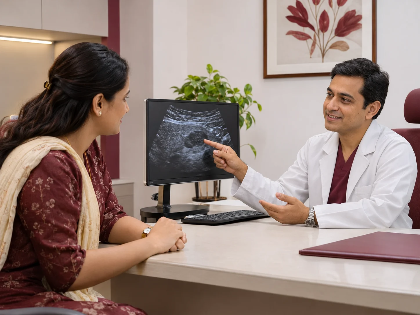
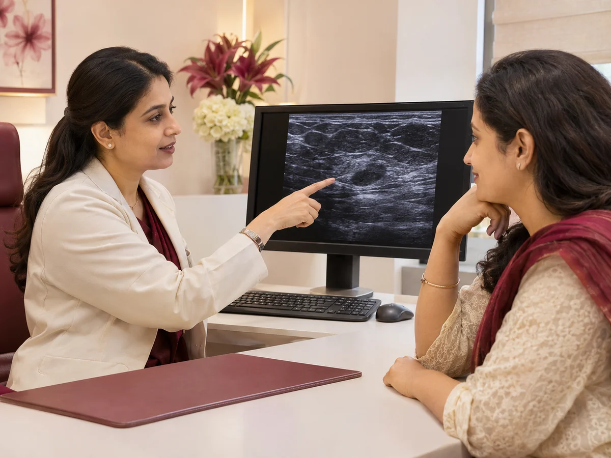
The imaging report and available ultrasound or mammography are reviewed, including the BI-RADS assessment when documented. We consider prior pathology, blood-thinning medicines, allergies and the need for mammographic rather than ultrasound guidance. Tissue results must be reviewed for agreement with the imaging findings, with breast-specialist follow-up as indicated.
What might treatment involve?
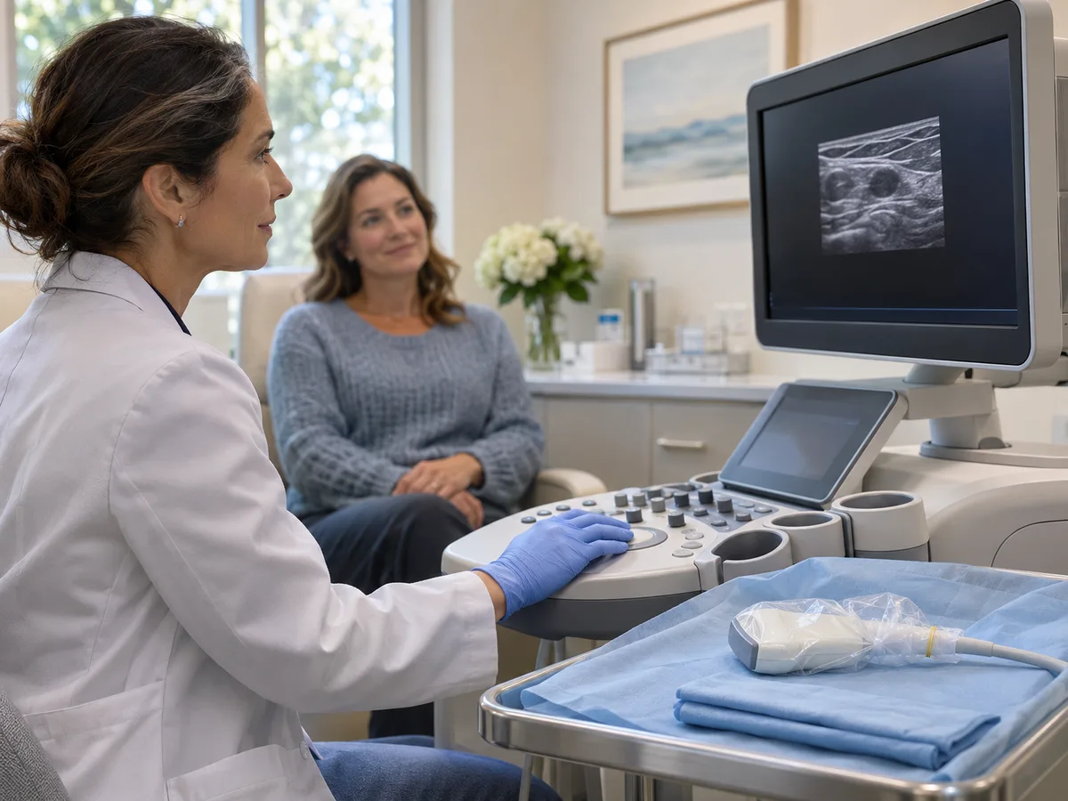
Depending on the imaging finding and treatment plan, a targeted ultrasound-guided procedure may be discussed. The exact technique, imaging method, laboratory arrangements and need for a breast surgeon are decided before scheduling.
Procedures that may be discussed
- Ultrasound-guided core needle biopsy: removes small tissue cores from a selected lesion for histopathology.
- Vacuum-assisted breast biopsy (VABB): uses a vacuum-assisted needle device to collect multiple tissue samples; selected benign lesions may be suitable for vacuum-assisted removal after appropriate evaluation.
- Breast cyst aspiration or selected collection drainage: removes fluid when clinically appropriate; suspicious findings may still require biopsy.
- Biopsy marker-clip placement: marks a sampled site for future imaging or treatment planning when indicated.
- Image-guided wire localisation: places a guide wire to assist the breast surgeon in finding a lesion for planned excision.
- Ablation of selected benign breast lesions: a separate specialist consideration after adequate diagnostic confirmation, with technique-specific eligibility and follow-up; it is not routine cancer treatment.
What else could help?
Imaging follow-up, a different biopsy method such as stereotactic or MRI-guided sampling, surgical biopsy or excision, or breast-specialist consultation may be preferable.
Risks and limitations
Pain, bruising, bleeding, infection, tissue injury, non-diagnostic sampling, marker displacement or the need for repeat biopsy can occur. A benign result that does not match suspicious imaging requires further assessment. Localisation is part of a planned surgical pathway, not the surgery itself.
Questions patients ask
Is a breast lump always cancer?
No. Many findings are benign, but imaging and sometimes biopsy are needed to establish a diagnosis.
Is VABB the same as breast surgery?
No. Vacuum-assisted biopsy samples tissue through a needle; selected lesions may be removed with the device, but suitability and complete removal cannot be assumed.
Why might a clip be placed after biopsy?
A small marker can help identify the sampled area during future imaging or treatment planning.
Does wire localisation remove the lump?
No. It helps the surgeon locate the area that is to be removed.